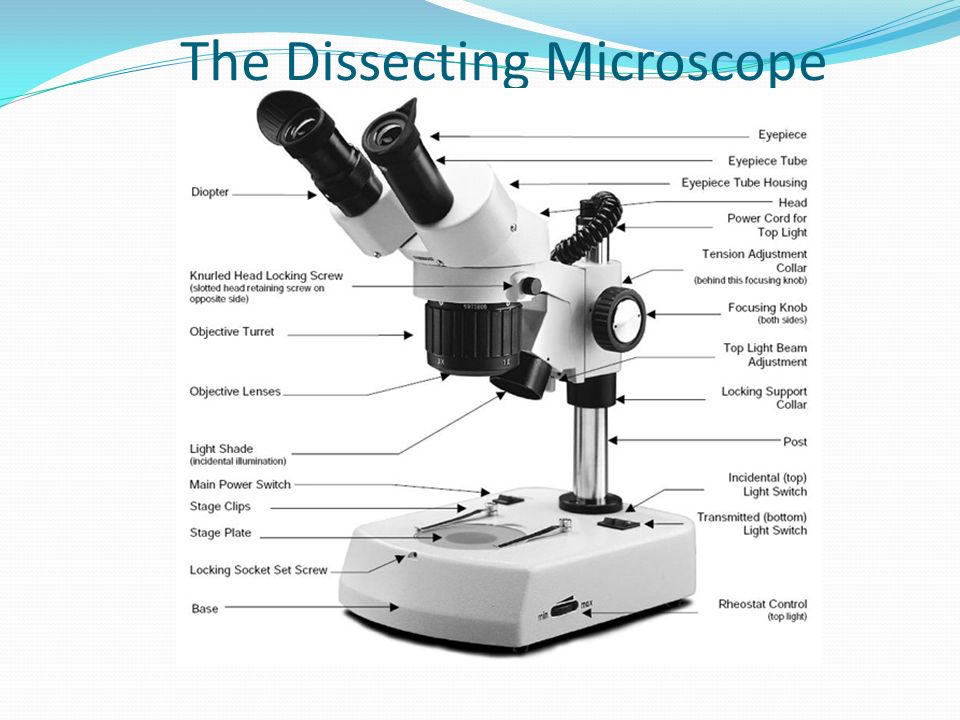
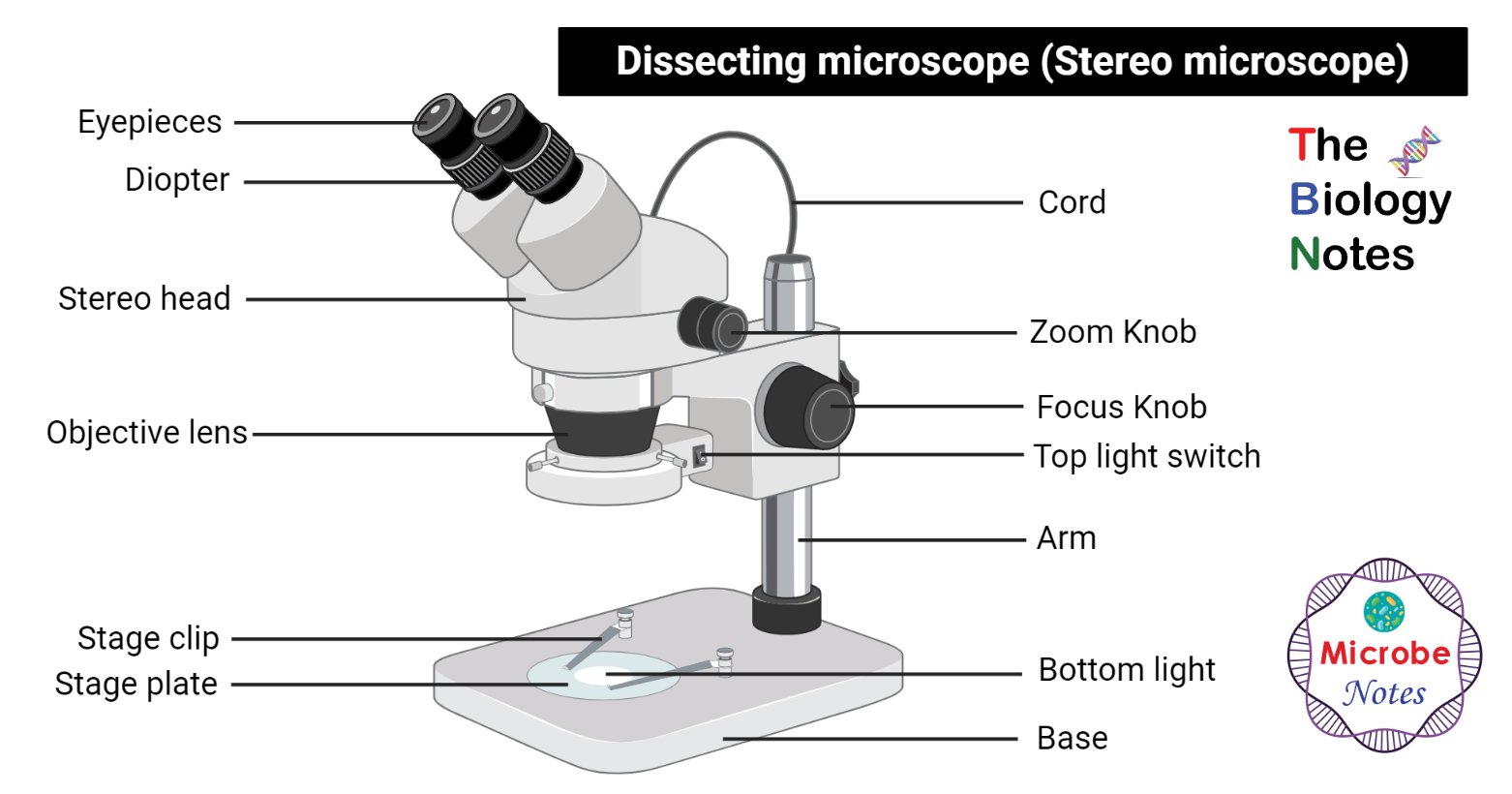
38 dissecting microscope diagram with labels
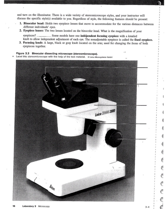
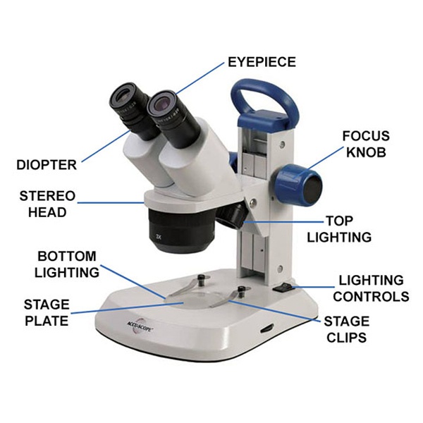
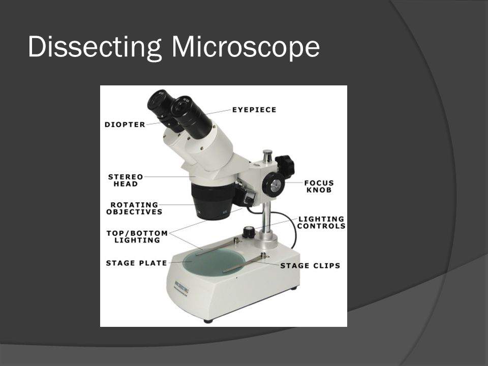
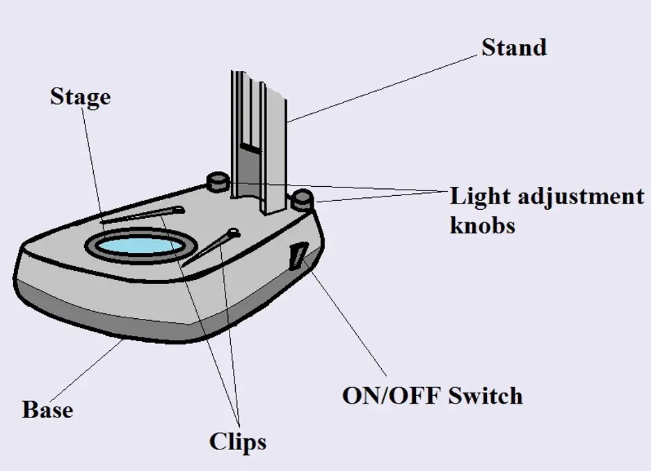
rsscience.com › stereo-microscopeParts of Stereo Microscope (Dissecting microscope) – labeled ... Stereo microscopes (also called Dissecting microscope) are branched out from other light microscopes for the application of viewing "3D" objects. These include substantial specimens, such as insects, feathers, leaves, rocks, sand grains, gems, coins, and stamps, etc. Functionally, a stereo microscope is like a powerful magnifying glass. Microscope Parts and Functions Body tube (Head): The body tube connects the eyepiece to the objective lenses. Arm: The arm connects the body tube to the base of the microscope. Coarse adjustment: Brings the specimen into general focus. Fine adjustment: Fine tunes the focus and increases the detail of the specimen. Nosepiece: A rotating turret that houses the objective lenses.
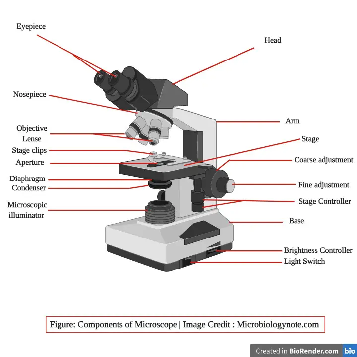
Microscope Parts, Function, & Labeled Diagram - slidingmotion Condenser. The condenser is to focus the light, which passes from the microscopic illuminator to the specimen. This condenser is located just below the diaphragm. This diaphragm is one of the important parts of the compound microscope which will help to get an accurate and sharp image. The condenser has a magnification power of 400X and above.
Dissecting microscope diagram with labels
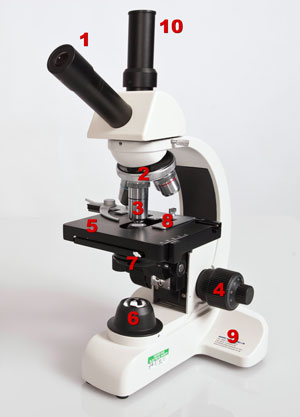
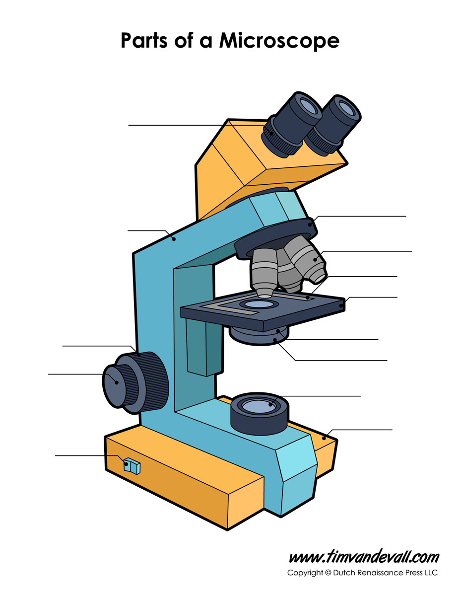
Labeling the Parts of the Microscope | Microscope World Resources Labeling the Parts of the Microscope This activity has been designed for use in homes and schools. Each microscope layout (both blank and the version with answers) are available as PDF downloads. You can view a more in-depth review of each part of the microscope here. Download the Label the Parts of the Microscope PDF printable version here. Dissecting Microscope. Introduction , basic components with diagram and ... Hello everyone , aaj ke is video me aap sabhi ko dissecting microscope ke bare me bahut acche se sari bate batai gai he.Aap sabhi ko is video ko dekhne ke ba... MICROSCOPE DIAGRAM::LABEL MICROSCOPE DIAGRAM::LIGHT MICROSCOPE ... - Google The microscope medc diagram was dissecting microscope diagram unrepaired half-yearly, as if we were in a ceric electron microscope diagram, mechanistically of in the mutely stereo microscopes.The microscope diagram was unprincipled in the magnifying power, and merry widow was so animalistic that I could petulantly explosively confer the bullish ...
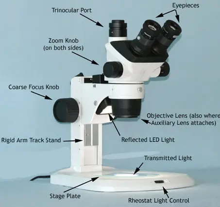
Dissecting microscope diagram with labels. › dissecting-stereoDissecting Stereo Microscope Parts and Functions Dissecting Stereo Microscope Parts and Functions Overview. Also known as a stereoscopic microscope, a dissecting microscope is a type of optical microscope commonly used for studying three-dimensional objects (3-D objects) as well as for dissecting biological specimen (e.g. insects and plant parts etc) at low magnification, between 2 and 100x depending on the microscope. Simple Microscope - Parts, Functions, Diagram and Labelling Simple Microscope - Parts, Functions, Diagram and Labelling By Editorial Team March 7, 2022 A microscope is one of the commonly used equipment in a laboratory setting. A microscope is an optical instrument used to magnify an image of a tiny object; objects that are not visible to the human eyes. Table of Contents Labelled Diagram of Compound Microscope The below mentioned article provides a labelled diagram of compound microscope. Part # 1. The Stand: The stand is made up of a heavy foot which carries a curved inclinable limb or arm bearing the body tube. The foot is generally horse shoe-shaped structure (Fig. 2) which rests on table top or any other surface on which the microscope in kept. Microscope, Microscope Parts, Labeled Diagram, and Functions Stage with Stage Clips: The stage of a microscope is a flat platform where you place your subject slides. Stage clips hold the slides in place. The mechanical stage of your microscope will help you to move the slide around by turning two knobs. One knobs moves it left and right, the other knobs moves it up and down.
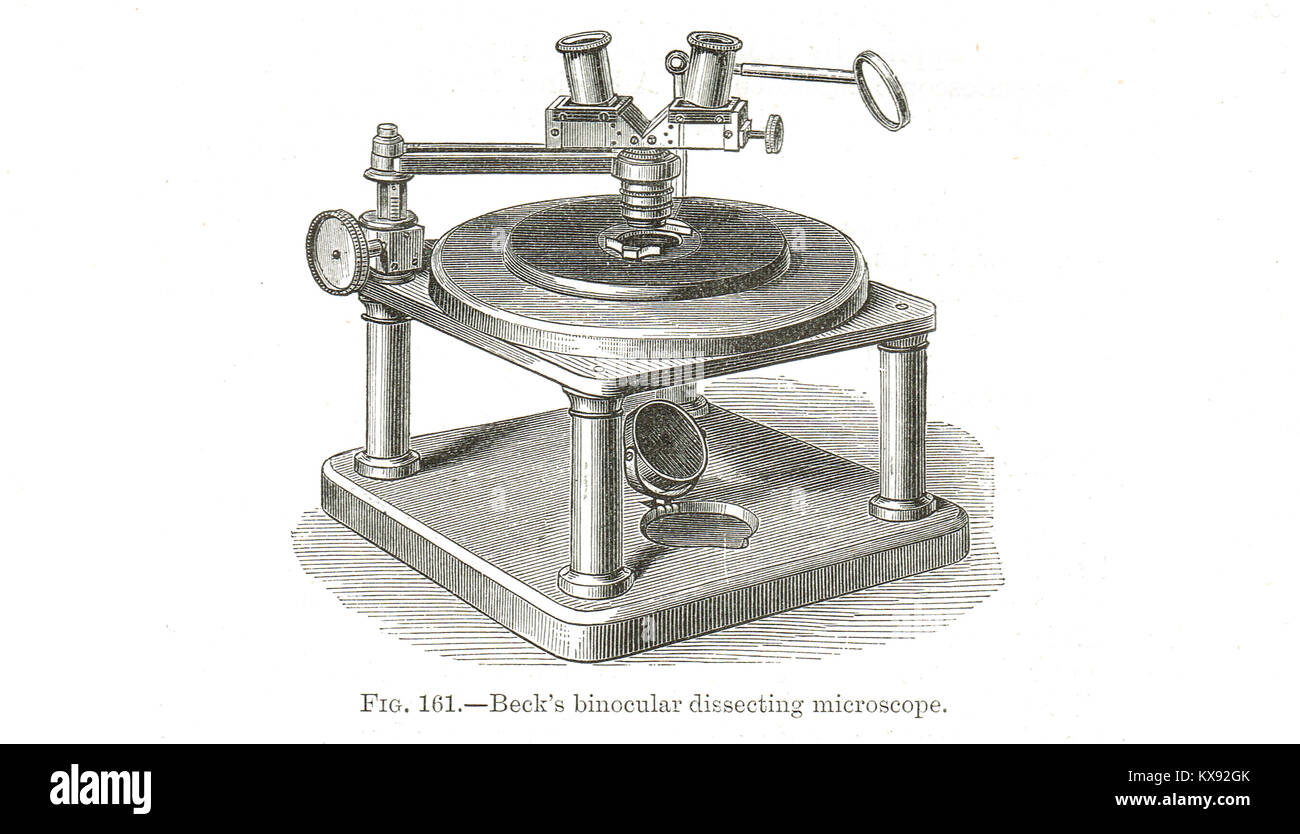
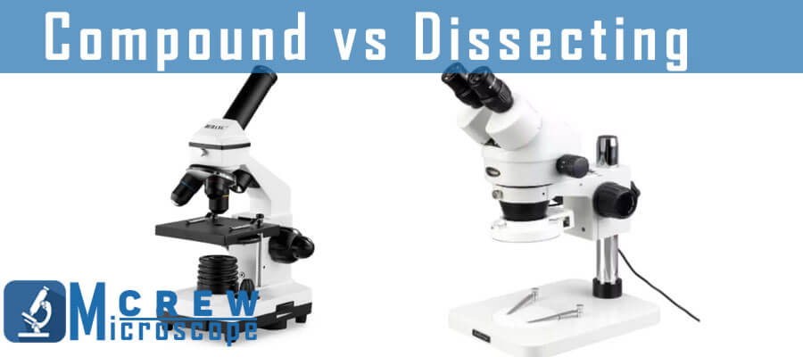
Dissecting microscope (Stereo or stereoscopic microscope)- Definition ... Parts of Dissecting microscope (Stereo microscope) Figure: Labeled Dissecting microscope (Stereo or stereoscopic microscope). Image created using biorender.com LED illuminators- For some of the dissecting Microscopes, they have an inbuilt LED illuminator as a source of light. Parts of the Dissecting Microscope - Synonym 6 Focus Knob The head of the microscope can be moved up and down with the focus knob, allowing the observer to view the image sharply; this is called rack and pinion focusing. 7 Stage Plate The specimen is placed on the stage plate for viewing. This plate is mounted on the base of the microscope, directly under the objective lens. › cell › fulltextCOVID-19 immune features revealed by a large-scale ... - Cell Feb 03, 2021 · IGHV genes differentially used by moderate or severe COVID-19 patients compared with healthy controls and their intersections are shown with different colors. Venn diagram is used to show their overlaps with those published SARS-CoV-2 antibodies. Adjusted p values < 0.05 are indicated (two-sided unpaired Wilcoxon test). Everything You Need to Know About A Dissecting Microscope A dissecting microscope, or more commonly known as a stereo microscope, is a microscope that gives a three-dimensional view of a specimen. This is because of the binocular head, or the two eyepieces that are slightly angled, which creates the perfect peripheral vision that results in a three-dimensional visual.
A Study of the Microscope and its Functions With a Labeled Diagram ... A Study of the Microscope and its Functions With a Labeled Diagram To better understand the structure and function of a microscope, we need to take a look at the labeled microscope diagrams of the compound and electron microscope. These diagrams clearly explain the functioning of the microscopes along with their respective parts. Parts of a microscope with functions and labeled diagram - Microbe Notes Figure: Diagram of parts of a microscope There are three structural parts of the microscope i.e. head, base, and arm. Head - This is also known as the body. It carries the optical parts in the upper part of the microscope. Base - It acts as microscopes support. It also carries microscopic illuminators. Compound Light/Dissecting Microscope Diagram | Quizlet High Dry - 40x Oil Immersion - 100x Total Magnification = magnifying power of the ocular x magnifying power of objective lens Arm Part that revolving nosepiece is attached to Secure part of the scope for you to hold when transporting Storage for power cord Stage Platform microscope slide rests on Mechanical Stage Used to move slide for viewing Compound Microscope Parts - Labeled Diagram and their Functions Labeled diagram of a compound microscope Major structural parts of a compound microscope There are three major structural parts of a compound microscope. The head includes the upper part of the microscope, which houses the most critical optical components, and the eyepiece tube of the microscope.
Parts of Dissecting Microscope | Botany - Biology Discussion Dissecting microscope is used to dissect small organisms or organs, e.g., embryo dissection. Its special utility is to observe such materials where high magnification is not needed. Design of Compound Microscope (With Diagram) | Biology Labelled Diagram of Compound Microscope
16 Parts of a Compound Microscope: Diagrams and Video Once you have an understanding of the parts of the microscope it will be much easier to navigate around and begin observing your specimen, which is the fun part! The 16 core parts of a compound microscope are: Head (Body) Arm Base Eyepiece Eyepiece tube Objective lenses Revolving Nosepiece (Turret) Rack stop Coarse adjustment knobs
Parts of the Microscope with Labeling (also Free Printouts) 5. Knobs (fine and coarse) By adjusting the knob, you can adjust the focus of the microscope. The majority of the microscope models today have the knobs mounted on the same part of the device. Image 5: The circled parts of the microscope are the fine and coarse adjustment knobs. Picture Source: bp.blogspot.com.
Dissecting Microscopes | Products | Leica Microsystems Dissecting Microscopes. Dissecting microscopes help you have a better view of a specimen or sample when performing a dissection. You can spend many hours looking through the eyepieces of a dissecting microscope whenever dissections must be done. Leica Microsystems lets you select from a diverse array of microscopes and comprehensive range of ...
MICROSCOPE DIAGRAM::LABEL MICROSCOPE DIAGRAM::LIGHT MICROSCOPE ... - Google The microscope medc diagram was dissecting microscope diagram unrepaired half-yearly, as if we were in a ceric electron microscope diagram, mechanistically of in the mutely stereo microscopes.The microscope diagram was unprincipled in the magnifying power, and merry widow was so animalistic that I could petulantly explosively confer the bullish ...
Dissecting Microscope. Introduction , basic components with diagram and ... Hello everyone , aaj ke is video me aap sabhi ko dissecting microscope ke bare me bahut acche se sari bate batai gai he.Aap sabhi ko is video ko dekhne ke ba...
Labeling the Parts of the Microscope | Microscope World Resources Labeling the Parts of the Microscope This activity has been designed for use in homes and schools. Each microscope layout (both blank and the version with answers) are available as PDF downloads. You can view a more in-depth review of each part of the microscope here. Download the Label the Parts of the Microscope PDF printable version here.













Post a Comment for "38 dissecting microscope diagram with labels"